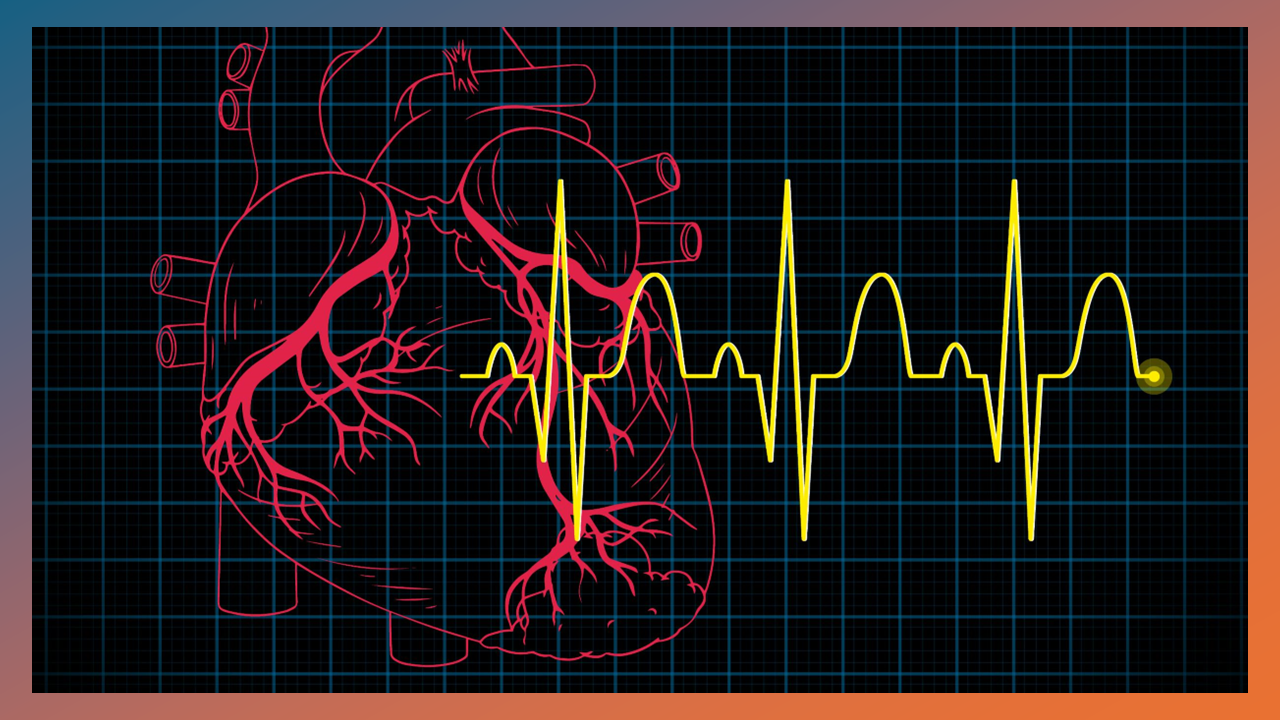
Why is your
"Heart" hard to Image, In Vivo ?
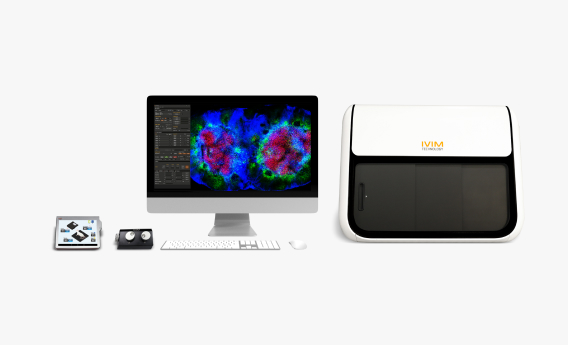
In vivo heart imaging is challenging because the heart is constantly moving with heartbeat and respiration, making stable, high-resolution imaging difficult. However, overcoming this enables real-time visualization of cardiac dynamics in a native physiological environment. To address this, we developed the engineered motion-stabilization solution specifically designed for stable in vivo heart imaging. |
|
|
Specialized Accessory ONLY for Heart Imaging |
|
|
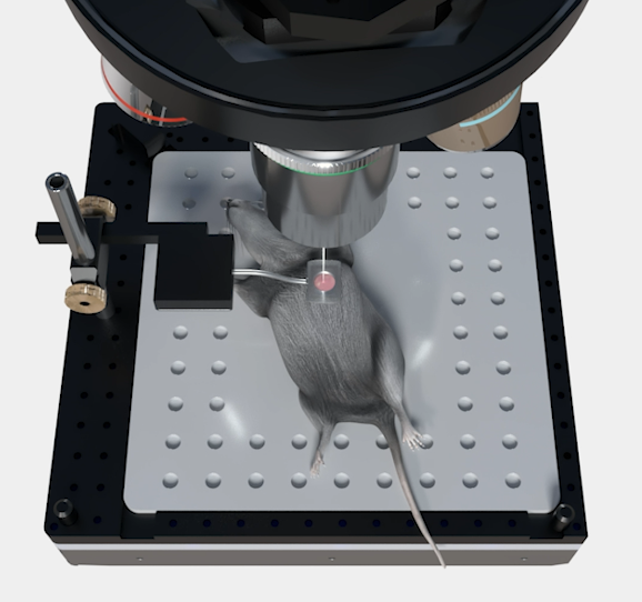
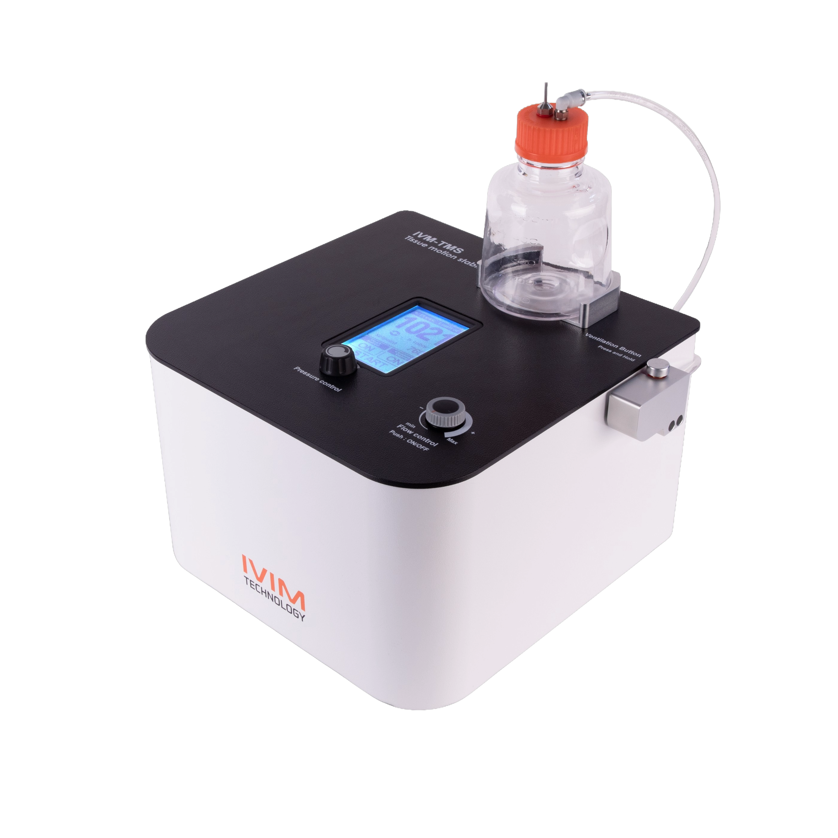
Tissue Motion Stabilizer (IVM-TMS)
Using IVM-TMS, negative pressure temporarily seals the space between the thoracic cavity and the imaging chamber, stabilizing the beating heart and for sequential in vivo imaging.
|
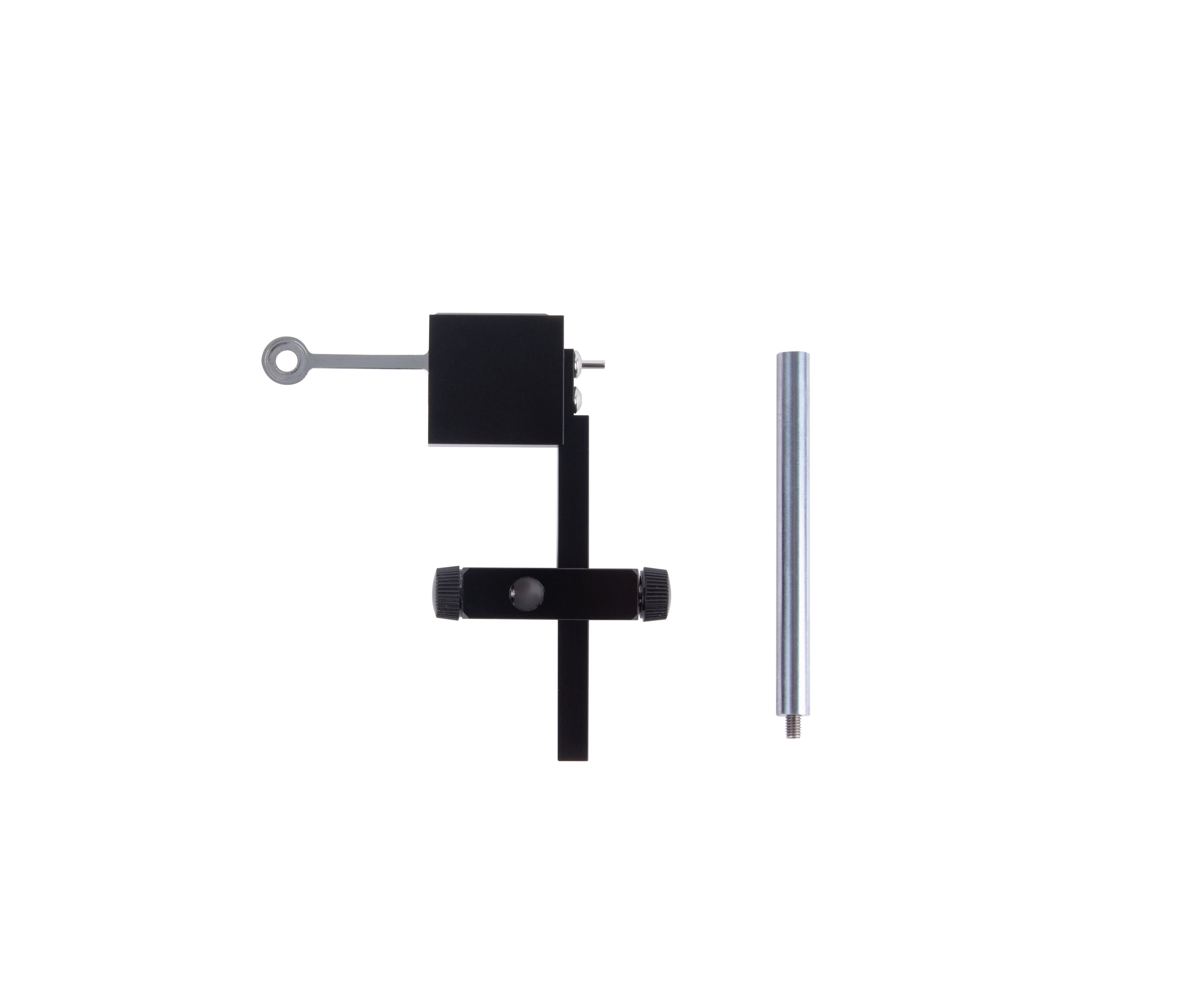
Vacuum-Based Chamber
The Vacuum-Based Chamber uses micro-suction technology with IVM-TMS to maintain negative pressure between the tissue and cover glass, minimizing micron-level motion in anesthetized mice. |
|
|
Additional Features to Improve Image Clarity! |
|
|
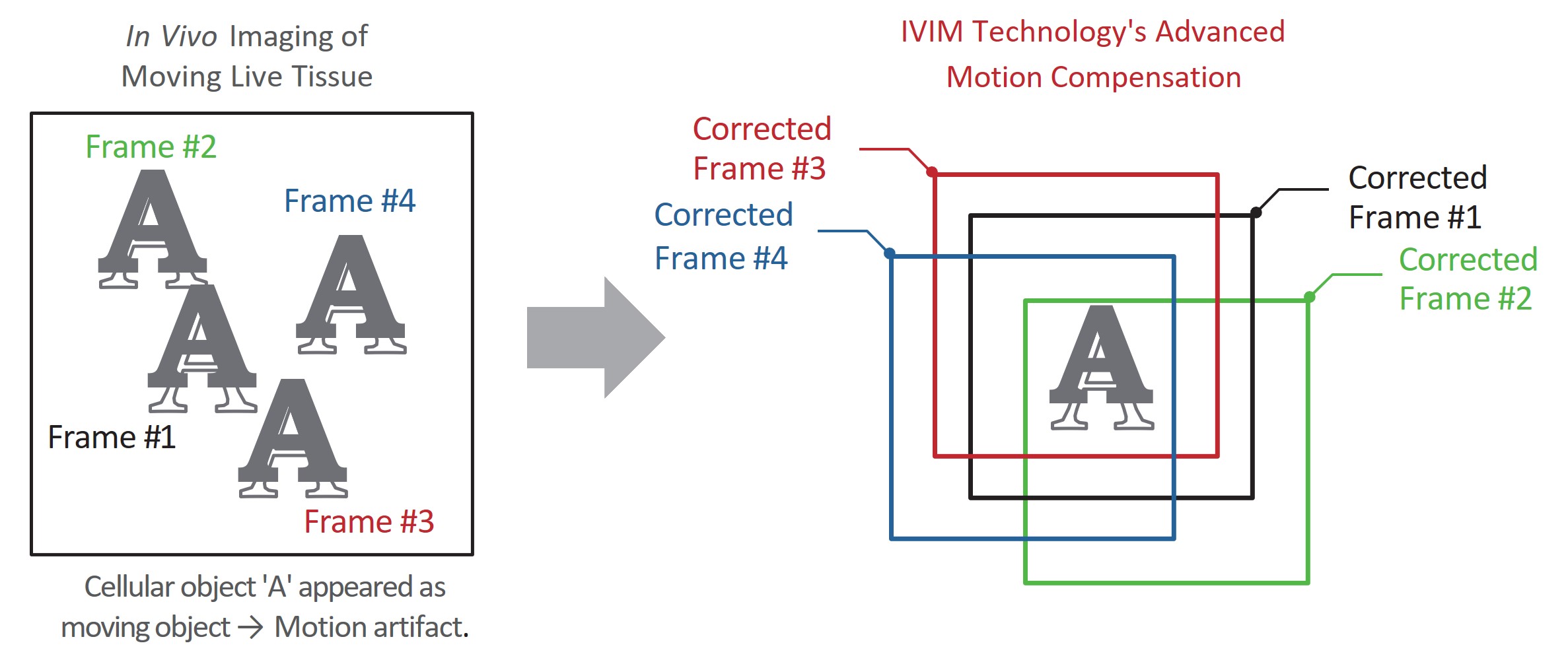
Principle
IVIM Technology provides one-click, high-precision motion compensation to correct respiration and heartbeat-induced movements during in vivo imaging. Using real-time video analysis, the algorithm automatically selects the least distorted reference frame and performs sub-pixel frame-by-frame correction, generating clear, high-quality images with GPU-accelerated processing.
Features
-
Motion Compensation: Automatic, hassle-free sub-pixel stabilization
-
Fast Processing: GPU-accelerated motion-corrected results
-
Spatiotemporal Support: Works across diverse tissue motions
-
Synergy: Optimized for ultrafast intravital imaging
|
|
|
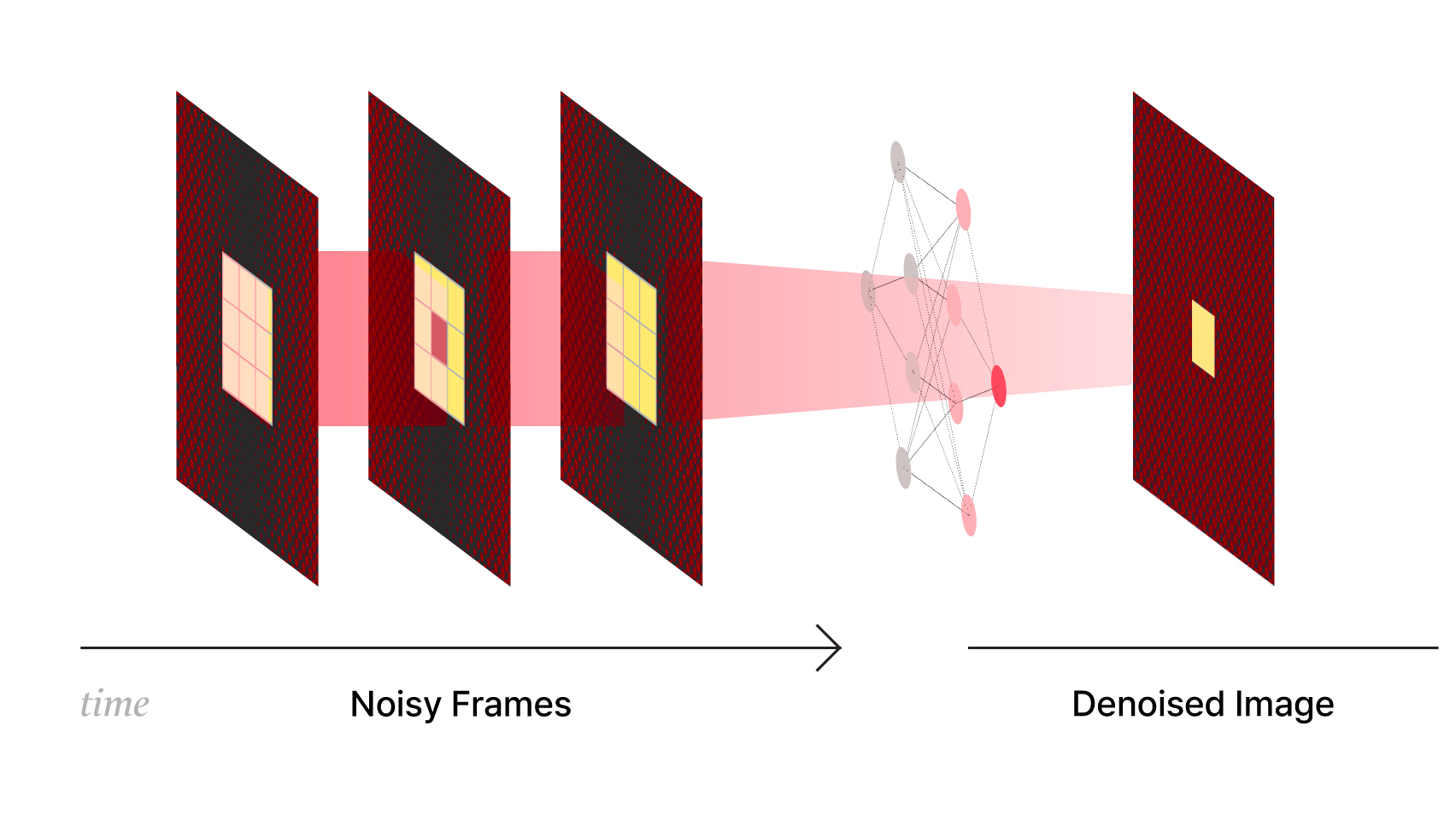
Principle
Dynamic organs like the heart are difficult to image because constant motion reduces SNR and obscures fine, fast biological events. Simply increasing imaging speed can further compromise image quality and timing precision. To address this, IVIM Technology adopts SUPPORT (Statistically Unbiased Prediction Using Spatiotemporal Information), a self-supervised denoising method that leverages spatiotemporal redundancy to remove Poisson–Gaussian noise while preserving fast dynamics for stable high-speed in vivo imaging. |
|
|
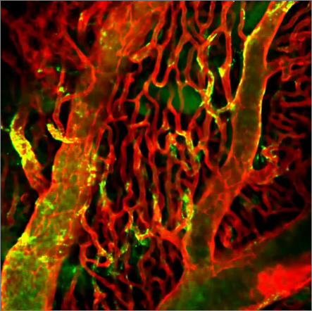
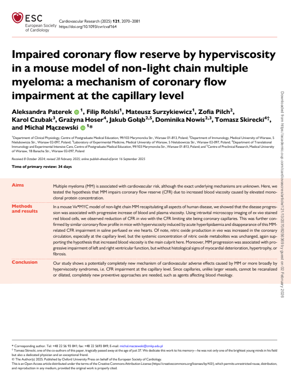
Recognized Among the Top 10
Cardio-Oncology Papers of 2025 |
|
|
This study, recognized among the Top 10 cardio-oncology papers of 2025 by the European Heart Journal, revealed hyperviscosity-driven capillary coronary flow impairment in multiple myeloma. Using IVIM’s in vivo Heart imaging, the team achieved real-time visualization of cardiac microvascular dynamics, highlighting how advanced in vivo imaging enables mechanistic insight in complex disease. |
|
|
2026 Special Event:
in vivo Heart Imaging Virtual Demo (Live) |
|
|
Virtual Demo Session 1
- March 5th │10:00 AM (CET), 09:00 AM (GMT)
Virtual Demo Session 2
- March 12th │10:00 AM (EDT), 9:00 AM (CDT)
|
|
|
|