Closing 2025 with IVIM’s latest advances in in vivo imaging IVIM TECHNOLOGY
NEWSLETTER
Volume 9, December 2025 |
|
|
About IVIM Technology, Inc.
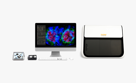
Founded in 2017, IVIM Technology is a global leader in turnkey intravital microscopy solutions. Our all-in-one box confocal and two-photon microscope systems enable real-time cellular imaging in live, anesthetized animals—transforming the landscape of in vivo research.
Recognized as one of the Top 10 APAC Bioanalytical Service Providers, IVIM serves a broad network of over 100 institutions worldwide, spanning academia, biotechnology, and the pharmaceutical industry. Our integrated portfolio includes intravital microscopy (IVM) systems, preclinical in vivo imaging solutions, training, specialized accessories, and labeled antibodies—empowering researchers to drive scientific discovery with precision, consistency, and efficiency.
|
|
|
In Vivo Imaging Apparatus:
The Reliable Path to Long-Term, Real-Time Observation |
|
|
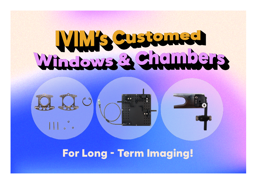
IVIM Technology’s intravital microscopy platform, combined with custom imaging windows and organ-specific chambers, enables longitudinal in vivo imaging—repeatedly visualizing the same tissue region in a single animal over days to weeks.
This approach minimizes inter-animal variability, increases statistical power, and reduces animal usage, while preserving the native tissue microenvironment.
By imaging identical regions over time, researchers can capture early and subtle biological changes, directly observe cellular and vascular responses to disease or therapy in real time, and reconstruct the temporal sequence of events that are often lost in end-point studies. |
|
|
Application 1 | Longitudinal Tracking of Tumor-Infiltrating T Cell Dynamics |
|
|
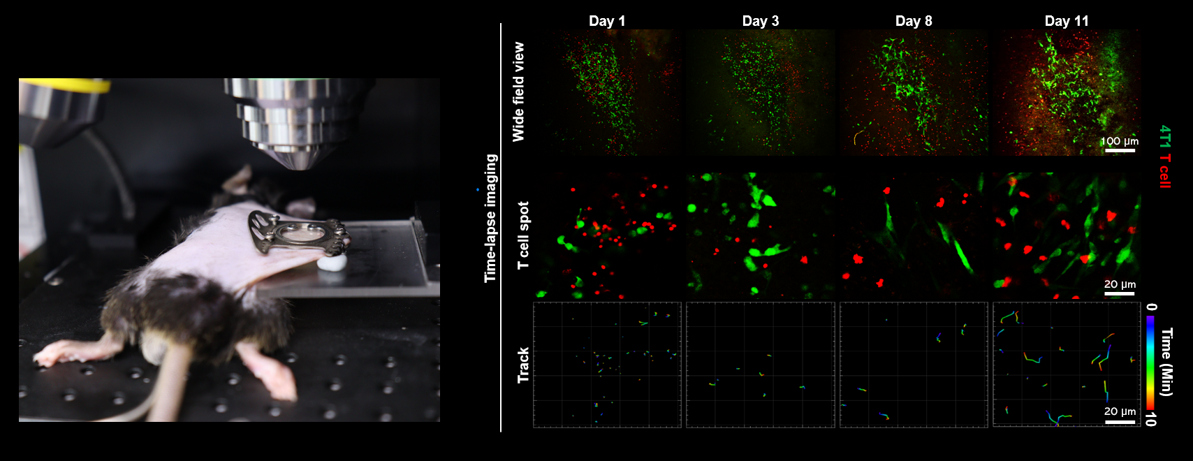
Figure 1. Longitudinal Imaging of Tumor-infiltrating Lymphocyte Movement in Breast Cancer Model
Window / Chamber: Dorsal Skinfold Chamber
The dorsal skinfold chamber enables stable, repeated access to the same tumor microenvironment over time. In this model, the same tumor region was longitudinally imaged at days 1, 3, 8, and 11 after implantation, allowing immune cell behavior to be tracked across early, intermediate, and later stages of tumor progression.
By imaging identical regions across more than a week, dynamic changes in T cell localization and motility could be directly quantified - revealing a gradual transition from early-stage immune quiescence to active T cell infiltration and migration within the tumor mass.
|
|
|
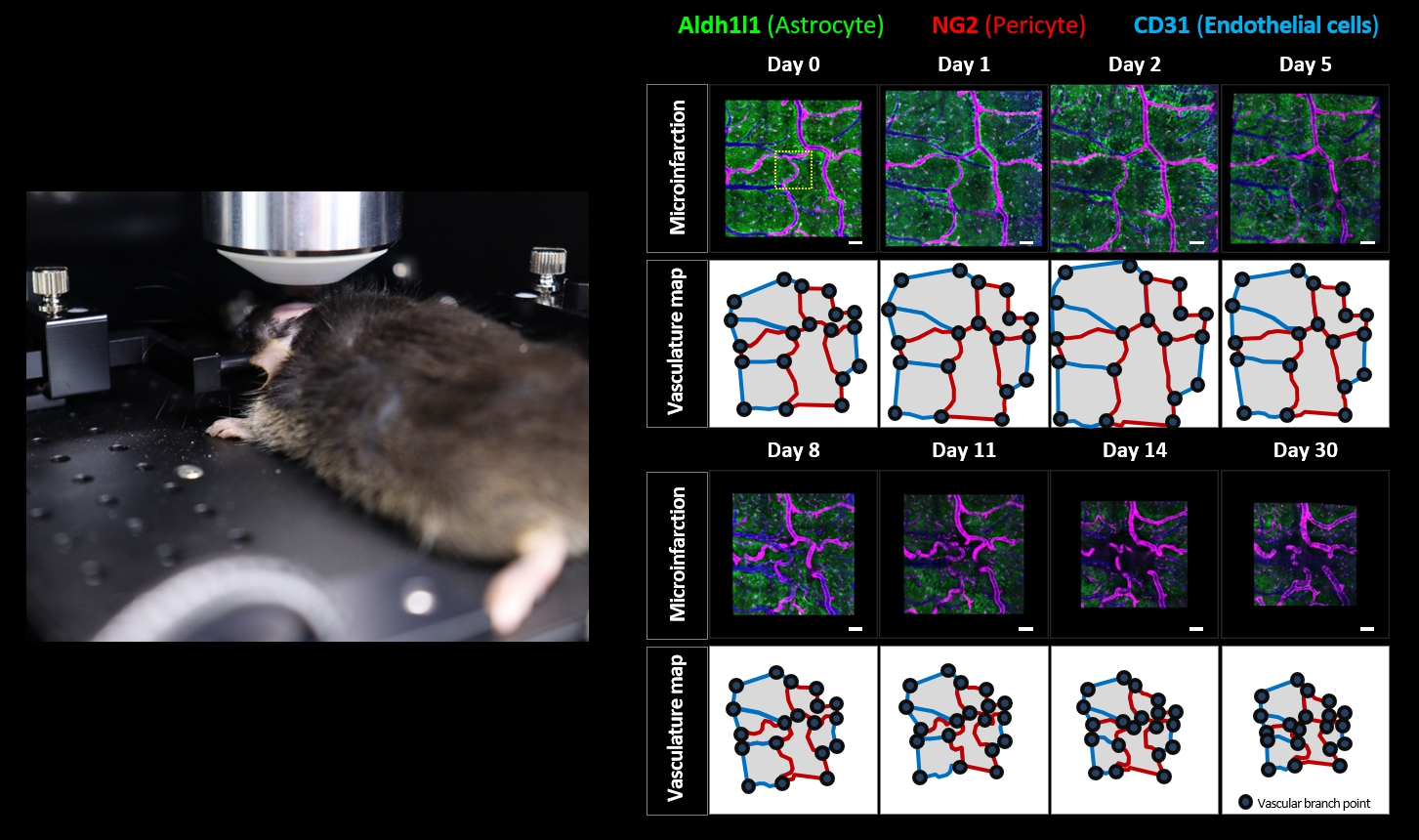
Application 2 | Mapping Neurovascular and Tissue Remodeling Over 30 Days After Microinfarction |
|
|
Figure 2. Longitudinal Imaging of Brain Shrinkage Followed by Photo-thrombosis in Cranial Imaging Window Model
Window / Chamber: Cranial Imaging Window
The cranial imaging window supports long-term, high-resolution imaging of cortical tissue and vasculature over weeks. Using this window, the same cortical region was repeatedly imaged at days 0, 1, 2, 5, 8, 11, 14, and 30 following targeted photo-thrombosis of a single arteriole.
This extended longitudinal timeline enabled direct visualization of the full progression - from acute vascular occlusion and edema in the first days, through gradual resolution, to delayed cortical shrinkage and chronic tissue remodeling over one month.
|
|
|
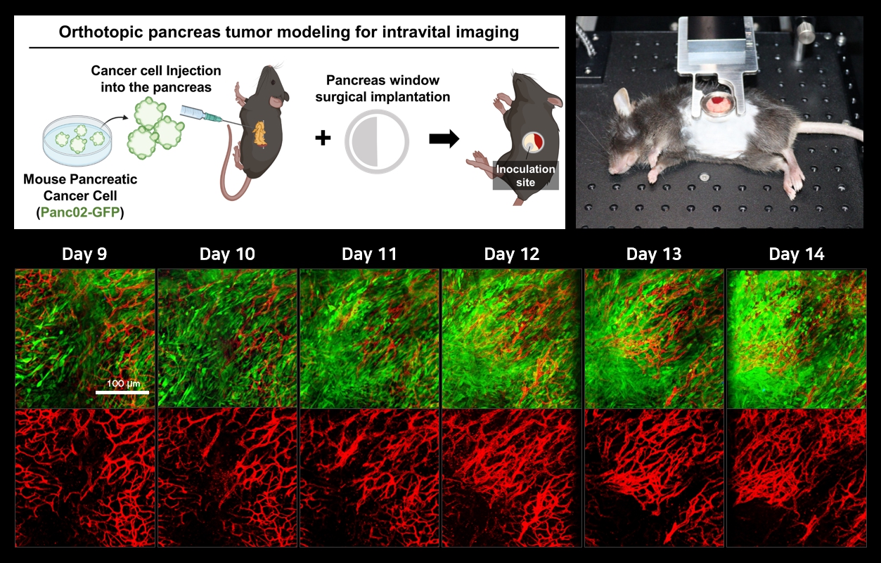
Application 3 | Monitoring Orthotopic Tumor Growth Over Multiple Days in a Deep Organ |
|
|
Figure 3. Longitudinal Imaging of Cancer Cell Growth in Pancreas Imaging Window Model
Window / Chamber: Pancreas Imaging Window
The pancreas is a highly dynamic and anatomically constrained organ, making repeated in vivo imaging particularly challenging. IVIM's pancreas imaging window overcomes these limitations by stabilizing pancreatic tissue and providing consistent optical access, enabling reliable longitudinal imaging of the same pancreatic region over time.
Using this window, orthotopic pancreatic tumors were repeatedly imaged from day 9 to day 14 after implantation, allowing continuous visualization of tumor growth together with angiogenic remodeling within the native pancreatic microenvironment without re-opening or re-positioning the tissue. |
|
|
Extending Long-Term Intravital Imaging Beyond These Applications!
Beyond the applications highlighted above, IVIM offers a wide range of organ-specific fixation adjuncts that support stable, long-term intravital imaging across diverse tissues and experimental settings. In addition, IVIM will participate in <REGENAGE 2026 – International Meeting on Ageing and Regenerative Medicine>, held in Singapore on January 26–27, where we will share insights and discussions focused on longitudinal in vivo studies. |
|
|
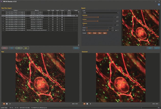
AI-Image Denoiser
IVIM’s AI-Image Denoiser improves low-SNR, high-speed imaging using self-supervised spatial-temporal AI, learning directly from raw data to preserve biological signals while reducing processing time and post-processing effort. |
|
|
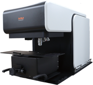
IVM-FS (Free Space)
IVM-FS is a free-space intravital microscopy system that expands in vivo imaging beyond mice to medium-sized animals such as rats, ferrets, and rabbits. Its open, stable design enables real-time confocal and two-photon imaging with motion compensation, supporting high quality, long-term imaging in physiologically relevant animal models. |
|
|
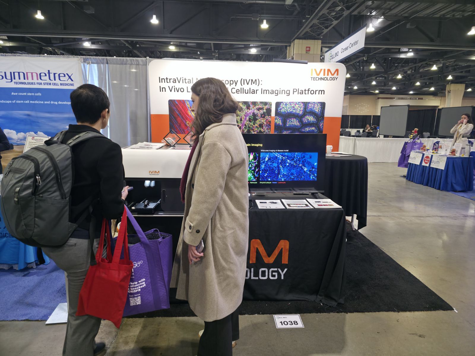
The Latest Event: Cell Bio 2025 | Philadelphia, PA |
|
|
IVIM Technology exhibited at Cell Bio 2025 (ASCB–EMBO Joint Meeting) in Philadelphia, presenting our intravital microscopy platform at Booth #1038. We also shared new in vivo imaging data through a scientific poster, highlighting real-time visualization of cellular dynamics in living systems. |
|
|
✉️ Year-End Message
As we come to the close of 2025, IVIM Technology advanced in vivo imaging technologies while strengthening its presence in the global research community through active participation in leading scientific conferences worldwide.
Looking ahead to 2026, we look forward to sharing more creative and innovative technologies, products, and research insights and we invite you to stay connected through our upcoming publications and events! Thank you. |
|
|
2025 Highlights
- Jan 21-23 | Advanced Imaging Methods (AIM)
- Mar 13-15 | BIO CHINA
- Mar 17-19 | Anatomy Physiology Pharmacology Week (APPW)
- Apr 25-30 | American Association of Cancer Research (AACR)
- May 3-7 | American Association of Immunology (AAI)
- Jun 16-19 | BIO 2025
- Jul 24-27 | Japan Neuroscience Society (JNS)
- Aug 17-22 | International Congress of Immunology (IUIS)
- Nov 1-2 | Japanese Vascular Biology and Medicine Organization (JVBMO)
- Nov 5-9 | Society of Immunotherapy of Cancer (SITC)
- DEC 6-10 | Cell Bio 2025
|
2026 in Focus
- Jan 12-15 | Fluorescence Advanced Imaging Research (FLAIR) 2026
- Jan 26-27 | REGENAGE Singapore 2026
- Apr 15-19 | American Association of Immunology (AAI)
- Jul 30-Aug 2 | Japan Neuroscience Society (JNS) Neuro 2026
- Sep 6-10 | International Vascular Biology Meeting (IVBM) 2026
- Nov 14-18 | Society for Neuroscience (SFN) 2026
- Nov 30-Dec 3 | British Society for Immunology (BSI) Congress 2026
... and more events ahead! |
|
|
IVIM TECHNOLOGY. All Rights Reserved.
|
|
|
IVIM Technologyinformation@ivimtech.com#A-1305, Hyundai Knowledge Industry Center, 11, Beobwon-ro 11-gil, Songpa-gu, Seoul, 05836, Korea
Tel: +82-2-431-7450Unsubscribe |
|
|
|
|